
|
Jurij Kotar impactThe vast majority of the equipment we have built over many years in our lab has been thanks to Jurij Kotar, whose expertise covers electronics, optics, coding, design and machining, as well as physics! Over time, this has culminated in some fantastic, very robust platforms, particularly for live cell automated microscopy and for optical tweezing. A variety of things can help a live cell experiment, from temperature and gas control, to bespoke chambers - it's impossible to list all the devices that Jurij has built. His work and advice has also inspired others in the group to develop hardware and software - I'd like to shout out to Luigi Feriani who developed multi-DDM in his PhD, and to Samuel McDermott, Morten Kals and Filip Ayazi who all built useful instruments and software.Optical Tweezers
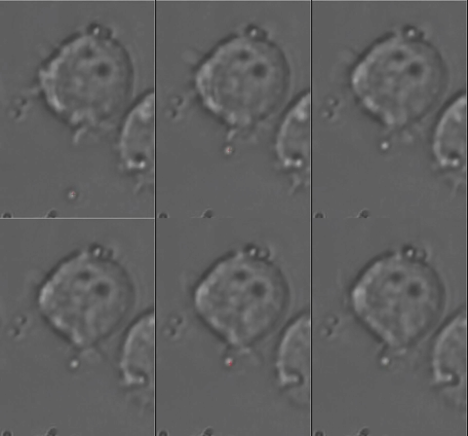
One of the key instruments developed by Jurij are optical tweeers (now in the 2nd generation). These are essential to our assays in malaria, and using them in closed-loop control has enabled unique lines of research in model cilia. Image and Video Analysis
A theme that spans across all our activities is the extraction of maximum information from image and video data. Microrheology is an example of this: the motion of tracer particles is tracked, and this information characterises the material in which the particles are embedded. Applications of this in our group are to study the mechanics of the cell cytoskeleton and the process of gelation in polymer and particulate systems. Instrument AutomationWe aim to automate as much as possible all our experiments, in order to improve data acquisition, and in some cases to enable experiments that it would simply not be possible to run manually. The optical tweezers and our imaging microscopes developed by Jurij can be run through a custom scripting language, which includes the possibility of feedback control via real time video analysis. These ideas have been developed in our lab also on simple cheap platforms like the OFM - see papers by Samuel McDermott.Microfluidics and Lab on ChipWe have built up facilities in our Physics Department, which now include two units for mask-less lithography (direct laser writing). The production of channels, pillars, structures in PDMS including multi-layer depths, is becoming routine. We can make micron scale incubators for continuous and controlled growth of bacteria, and we are developing devices to study other unicellular organisms. On this we have learned a lot from Kevin Dorfman at the University of Minnesota.Liquid interfacesWe maintain and still develop a suite of instruments for the measurement of liquid surface tensions, and interface shapes.Instruments and gadgets exploiting 3d printing - low costIt is amazing what can be prototyped with the current generation of 3d printers, and simple microcontrollers and stepper motors. Samuel McDermott led on a couple of very low cost objects, which function well and I hope will have impact: (1) an automated gadget to make blood smears, which makes the process demonstrably more reproducible. [S.McDermott et al., Rev. Sci. Instr. 93, 014104 2022] (2) a device to "retrofit" old lab equipment, and allow it to perform in an automated way (imagine a "scientific" smart plug). [S.McDermott et al., RSOS 11, 240634 2024] We have worked on these projects always as open source projects, with an emphasis on documentation to allow others to build or fork their own projects. We have a lot of projects in this space, at various stages of development. Further development can be fun for final year students, or summer projects, so get in touch if this seems your thing.
| |

 |
 |
 |